


You may have been told you need a scan for your knee, or wondered whether an X-ray or MRI would give a clearer answer. It can be confusing when different tests are mentioned, especially when the symptoms feel similar.
Not every case of knee pain requires advanced imaging. Orthopaedic surgeons decide which scan is appropriate based on your symptoms, physical examination and medical history. Understanding how these decisions are made helps explain why a particular test is recommended and what it means for your treatment.
What Causes Knee Pain and When Should You Seek Evaluation?
Knee pain can result from a wide range of conditions and affects individuals of all ages and activity levels. It may develop suddenly due to an injury or gradually over time from repeated strain or underlying joint changes.
Common causes include acute injuries such as ligament sprains, meniscus tears or fractures, often linked to sports and dance or accidents. Overuse injuries, such as tendonitis or runner’s knee, can occur from repetitive movements without adequate rest. In older adults, degenerative conditions like osteoarthritis are a frequent cause, leading to pain, stiffness and reduced mobility.
While mild discomfort may improve with rest and activity modification, certain symptoms should prompt medical evaluation. These include persistent pain, noticeable swelling, instability, locking of the knee or difficulty bearing weight. Pain that interferes with daily activities or does not improve over time should also be assessed.
X-Ray vs MRI: What’s the Difference?
When evaluating knee pain, X-rays and magnetic resonance imaging (MRI) scans are commonly used imaging tools, but they serve different purposes.
An X-ray is typically the first-line imaging test. It is quick, widely available and particularly useful for assessing bones and joint structures. X-rays can detect fractures, joint alignment issues and signs of osteoarthritis, such as narrowing of the joint space or bone spurs.
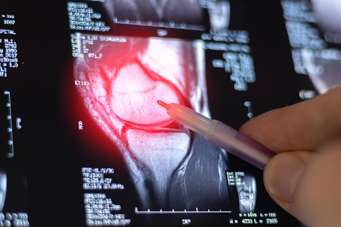
An MRI provides more detailed images of the soft tissues within the knee. This includes ligaments, tendons, cartilage and the meniscus. MRI scans are especially helpful in identifying injuries such as ligament tears, meniscus damage or other internal joint problems that may not be visible on an X-ray.
While MRI offers more detailed information, it is not always necessary as a first step. X-rays are often sufficient for initial assessment, with MRI reserved for cases where further evaluation is needed to confirm the diagnosis or guide treatment decisions.
How Do Orthopaedic Surgeons Decide Which Scan You Need?
Choosing between an X-ray and an MRI is not a one-size-fits-all decision. Orthopaedic surgeons base their recommendations on a combination of your symptoms, how the injury occurred and findings from a physical examination.
Medical history and symptoms
Your doctor will assess when and how the pain started, along with symptoms such as swelling, instability, locking or clicking. They will also consider how the condition affects your daily activities or sports.
Physical examination
A clinical examination helps evaluate your range of motion, joint stability and areas of tenderness. Specific tests may be performed to identify ligament, meniscal or tendon injuries.
Based on these findings, the most appropriate imaging test is then selected to better understand the cause of your knee pain and guide treatment.
What Are the Next Steps After Imaging?
Imaging results are only one part of the overall assessment. Orthopaedic surgeons interpret scans alongside your symptoms and physical findings to form an accurate diagnosis and guide the next steps.
Interpreting your results
Findings from X-rays or MRI scans are reviewed in context, as not all abnormalities seen on imaging are the cause of the pain. Your doctor will explain what the results mean and how they relate to your symptoms.
Non-surgical treatment options
Many knee conditions can be managed conservatively with physiotherapy, medications or activity modification. These approaches aim to reduce pain, improve function and support recovery without surgery.
Further evaluation if needed
If the diagnosis remains unclear or symptoms persist, additional assessments or follow-up imaging may be recommended to better understand the condition.
Surgical considerations
Surgery may be advised for more severe injuries or when non-surgical treatments are not effective. The decision is based on the type of condition, severity and your activity goals.

Precise Diagnosis for Knee Pain at the Bone & Joint Centre
Knee pain can have many underlying causes and choosing the right imaging test is an important step in reaching an accurate diagnosis. Orthopaedic surgeons consider your symptoms, examination findings and individual needs before recommending an X-ray or MRI. Seeking timely evaluation ensures appropriate treatment and supports better long-term outcomes.
At The Bone & Joint Centre, we provide specialist evaluation for knee pain, ensuring that each patient receives the most appropriate imaging and treatment approach based on their condition. Patients are in the trusted care of Dr Kevin Koo Oon Thien, who completed a one-year fellowship under the Health Manpower Development Plan (HMDP) Award at St. Mary's and Charing Cross Hospitals, Imperial College Healthcare in London, UK. If you are experiencing knee pain and are unsure whether you need an X-ray or an MRI, schedule a consultation with us for a specialist-led management plan.